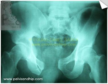
Anteroposterior view of a rotationally and vertically unstable fracture. The posterior failure is through the right SI joint with complete disruption of all components of the posterior SI ligament complex. Vertical displacement may not be very obvious but the vertical instability is evident.
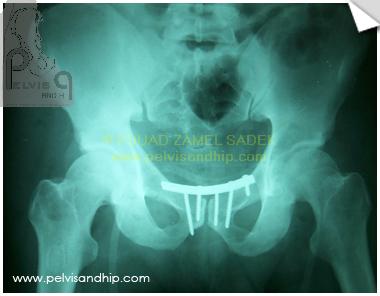
Anteroposterior view after the first surgery. The initial treating surgeon treated this injury as a rotationally unstable injury only. Fixation was done through a pfannestiel incision with stabilization of the symphysis only. The right SI joint seemed adequately reduced judging on the closed symphyseal gap only.
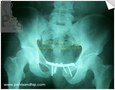
With follow up, the inadequate sole anterior ring fixation started to fail with secondary displacement of the pelvic injury with opening up of the symphysis and a starting vertical displacement of the right SI joint which did no show on the initial preoperative films. Notice the pulling out of the screws.
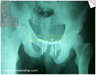
A more closer look to the outlet view shows the vertical displacement more. The failure of the symphyseal plate is also obvious.
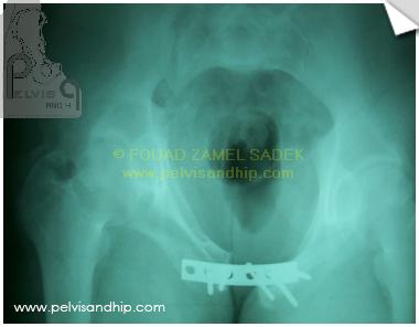
The inlet view showing more of the displacement with the posterior displacement at the level of the right SI joint obvious.
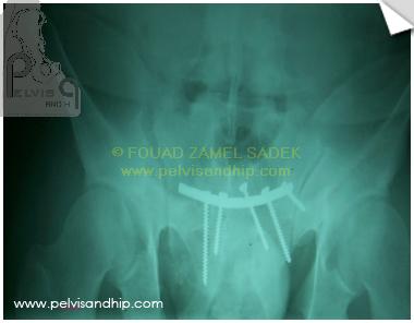
The outlet view showing the displacement.
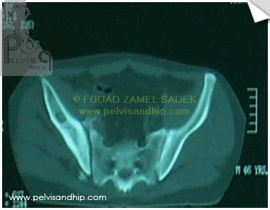
The axial CT scan of the pelvis showing the displacement of the SI joint with some evidence of callus formation at the margin of the SI joint.
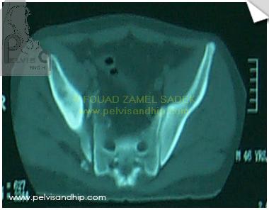
Opening up of the right SI joint more obvious at the inferior aspect of the joint.
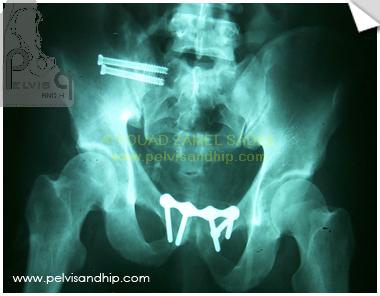
We operated upon him after more than 4 months of the primary surgery due to pain. A pfannestiel incision done for the removal of the anterior plate with refixation after reduction of the symphsis. An anterior SI approach was done to curett the joint and mobilize it prior to the attempt of reduction of the symphysis. Posterior fixation was done with two SI screws.
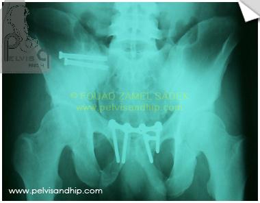
Outlet view showing the satisfactory reduction and fixation with one anterior symphyseal plate and 2 SI screws. Patient was fully mobile within 3 months with no further disability. He returned to work as a construction worker within 6 months.
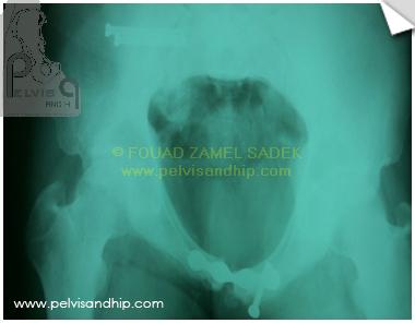
Inlet view showing reduction and fixation of the injury.

If you feel like posting comments, enquiries or questions please click here.