
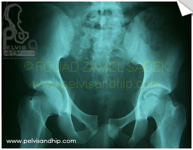
Rotationally and vertically unstable injury in a 19 years old male with signficant widening of the sacral fracture on the right side. A near internal hemipelvictomy injury on the right side.
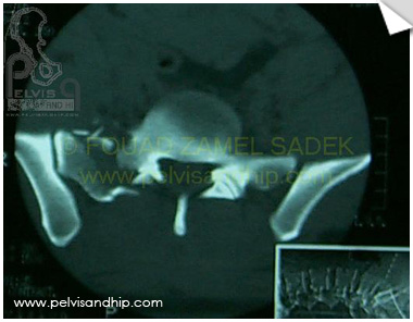
Axial CT scan show lateral sacral alar fracture on the right side with wide separation. Fracture is comminuted.
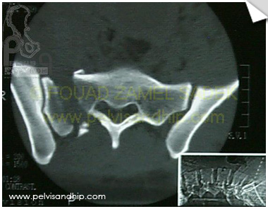
Gap is more widened as we go distally on the axial CT scan.
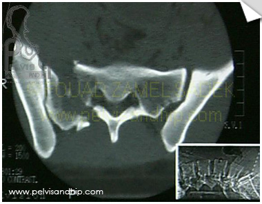
At the level of the sacral foramen the fracture is no through the neural canals minimizing risk in case of SI screw fixation.
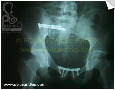
Operated upon after 3 days from the injury, through pfannestiel incision open reduction and internal fixation of the symphysis was done. This was followed by SI screw fixation with the patient in the prone position. Nowadays the second procedure is routinely done in the supine position.
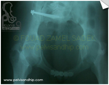
Inlet view shows the satisfactory ring recostruction with the 2 SI screws in the sacral bodies.
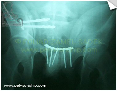
The Outlet view shows the reduction and fixation. On the pubic bodies most of the screws are adequately inserted gaining good purchase. Note that the lower SI screw is above the first sacral foramen. The upper SI screw is not in the most ideal direction being just below the lumbosacral trunk; also, it is slightly long into the disc. Luckily, patient had no clinical consequences with satisfactory function with no need for follow up removal.

If you feel like posting comments, enquiries or questions please click here.