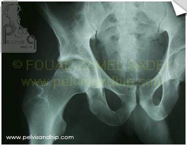
AP view pelvis of an 18 years old motorcyclist admitted to ER for an open fracture of the right tibia. He also had this apparently innocent injury of the right acetabulum; what looks like a posterior wall fracture with minimal displacement on this view. He had initial emergency management for the tibial fracture and was transferred to our institution for follow up of the tibia. Further studies of the acetabular fracture revealed the true nature of the injury.
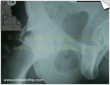
On the obturator oblique view one can identify a comminuted displaced fracture of the posterior wall. The anterior column seems intact.
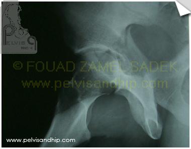
On the Iliac oblique view the posterior column is fractured with signficant displacement. The upper end of teh fracture can be easily depicted at the top area of the greater sciatic notch. The 3 views analysis identify an associated posterior wall posterior column.
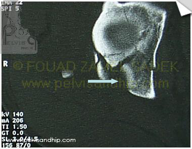
Further assessement revealed at the axial cut above the dome, the association of the posteior column fracture with a high posterior wall with marginal impaction (notice arrow)
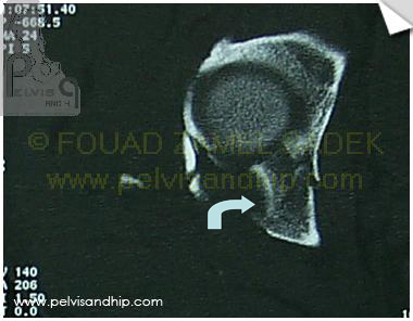
An axial cut CT at the level of the upper end of the femoral head show clearly the marginally impacted fragment which needs to be elevated and grafted prior to reduction of the posterior wall fragment (notice arrow)
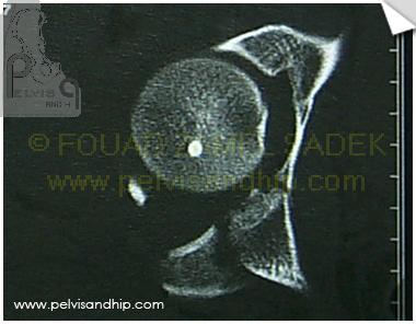
The posterior wall fragment clearly seen on this axial CT cut
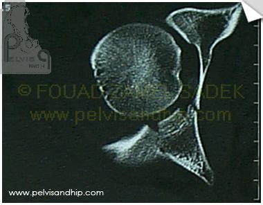
The inferior limit of the posterior wall as seen by the CT cut at the level of the ischial spine.
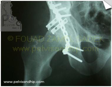
AP view of the hip after open reduction and internal fixation through a Kocker-Langenbeck approach. In these days we used to operate on these in the lateral position, nowadays we do that in the prone position. A 3.5 reconstruction plate is used for fixation of both posterior wall and column. A divided 1/3rd tubular plate is used for buttressing 2 comminuted fragments of the posterior wall.
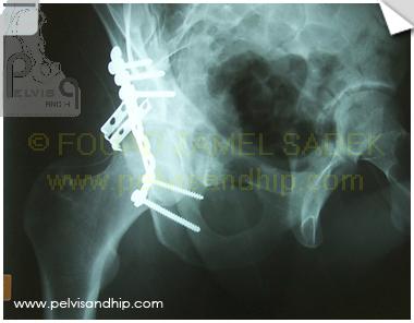
Obturator oblique view of the hip showing the well reduced posterior wall in profile with the 2 buttress plates supporting the comminuted posterior wall fragments.
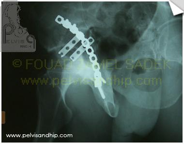
The iliac oblique view showing the coutoured reconstruction plate lying on the posterior column and the two buttress plates; what are sometimes called spring plates.
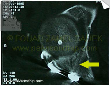
Satisfactory elevation, grafting and fixation of the marginally impacted fragment. Notice the place of the graft medial to posterior wall proper. (arrow)
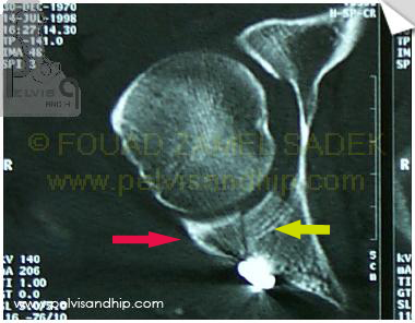
Axial CT cut showing the elevated and grafted marginally impacted fragment (yellow arrow) beside the reduced posterior wall proper (red arrow)
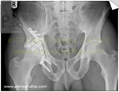
At 9 years follow up this hip is rated 18 by the Merle D’aubigne and Postel score.
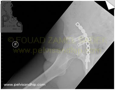
The iliac oblique view showing the hip at 9 years follow up with good preservation of the hip joint.
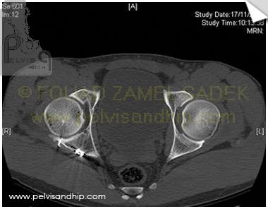
CT scan at 9 years follow up of the hip with appearance of the healed previously elevated and grafted marginally impacted fragment.

If you feel like posting comments, enquiries or questions please click here.