
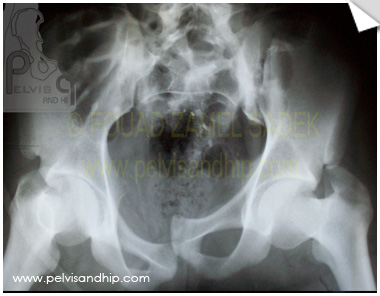
An antero posterior view of case 1 with a locked symphysis and a fracture dislocation of the left SI joint. Mild vertical displacement. A locked symphysis is an indication for reduction with or without fixation as per case.
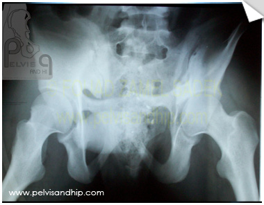
Outlet view showing clearly the overlapped symphysis with no disc appearance. On the outlet view the almost no vertical displacement can be depicted.
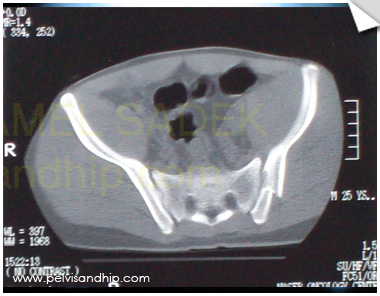
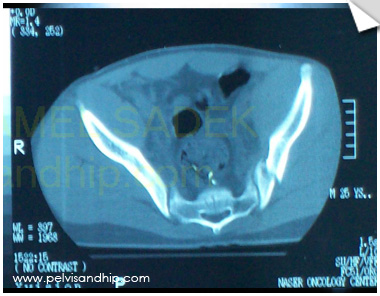
The fracture involving the left SI is very clear on this axial view of the pelvis. Such a fracture is more tricky to fix with an SI screw but quite amenable to anterior SI plating.
Another view showing the SI fracture dislocation with the limited path for the SI screw fixation.
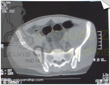
The fracture is involving the most inferior part of the symphysis.
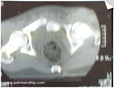
The CT axial cut at the level of the symphysis showing clearly the locked nature of that lesion.
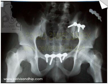
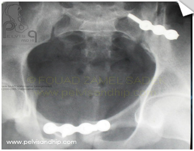
The postoperative AP view after open reduction and internal fixation of both lesions; the symphysis was reduced through a pfannestiel incision and one plate was deemed stable. An anterior SI approach was used for fixation of both SI joint and the iliac wing fracture extension.
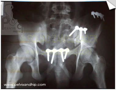
The near outlet view revealing the fixation. Note that in an ideal outlet view the pubic tubercle should be opposite the 2nd sacral foramen.
The closer to inlet view after reduction and fixation. Note that when a double plating construct is used the plate can be placed closer to the anterior margin of the SI joint to accomodate another plate posteriroly.

If you feel like posting comments, enquiries or questions please click here.