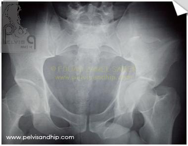
AP view of an associated both column fracture of the left aceabulum in a 29 years old male. Notice that the left SI joint is sligtly opened. On the AP view overshadows of the iliac extension of the fracture can be seen pointing to a floating acetabulum. The posterior column component is high. There is some central head subluxation. An anatomic reduction is mandatory in this case.
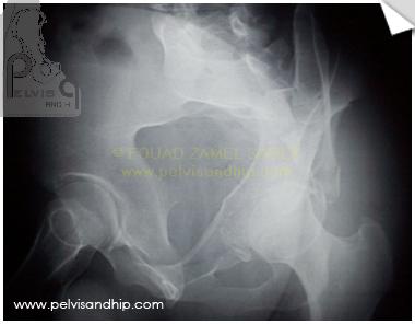
Obturator oblique view showing the central dislocation. The spur sign can be identified although it is a rather undisplaced spur due to inplace position of the superior wall component. There is some secondary fracture along the anterior column which requires special fixation for the quadrilateral plate component.
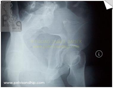
The iliac oblique view stresses the posterior column fracture which is high enough for an ilioischial screw. A faint shadow is standing vertically just medial to the femoral head (the quadrilateral plate).
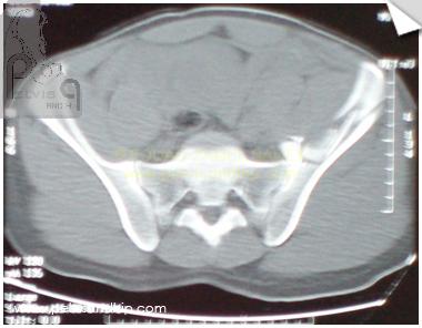
The value of axial CT cuts is to follow the different cuts in a serial way from proximally to distally. A cut at the upper part of the left SI joint showing the comminuted fracture nature of the ilium.
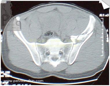
Going lower down the SI joint involvement in the high fracture of the ilium can be depicted. This does not mean that there is an associated pelvic ring disruption, but it has to be considered in the plan of fixation of the acetabular fracture.
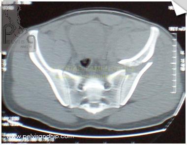
The inferior part of the SI joint is seen clearly with an intact SI joint ligaments.
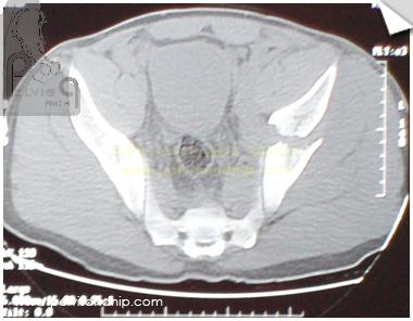
The anterior column fracture component can be identified in the coronal plane above the dome of the acetabulum.
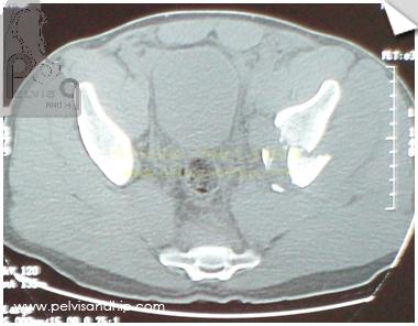
The 2 upper part of the anterior and posterior columns can be identified in this cut as 2 separate bony islands.
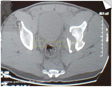
The quadrilateral plate will be more identified in this plane
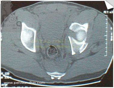
On this cut most of the quadrilateral plate seems to be attached to the posterior column. A small cortical flake of bone which could represent a small postero superior wall is present but it is not signficant enough to warrant a posterior approach.
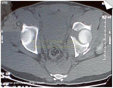
A more distal cut at the level of the upper part of the femoral head show the inward displacement of the head with gaping between the anterior and posterior columns.
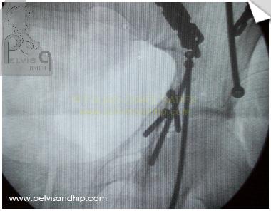
Through an ilioinguinal approach the fracture was reduced in a step like fashion, starting with the SI joint component first with fixation with a 3 hole reconstruction plate. The ilium was reduced and fixed with an interfragmentary screw with another buttress plate. This was followed by the quadrilateral plate which was fixed by the so-called wedge screw (the smallest and most distal screw along the anterior column on this image intensifier picture).
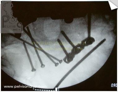
As the posterior column part of the quadrilateral plate was the bigger and more signficant part 3 long ilioischial screws were inserted from the brim into the posterior column (the screws between the plate on the right side and the last or smaller screw on the left). Notice that on this view one can clearly see the extraarticular pathway of the screws to avoid joint penetration (one can clearly see the femoral head on the right side with the short screw superimposed)
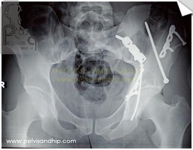
Postoperative AP view of the pelvis. The femoral head is adequately reduced. All lines are anatomically reduced and the pelvic ring is well formed. Notice bending of the long ilioischial screw which can happen with these long 3.5 screws.
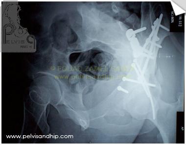
Obturator oblique view show the reduction and fixation. One can identify the small postero supreior fragment of bone which was left undisturbed; a posterior approach for this fragment was not felt to be warranted.
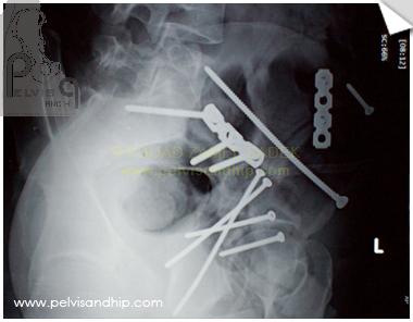
Postoperative iliac oblique view showing clearly the so-called LC II screws fixing the ilium. The 3 ilioischial screws into the posterior column can be easily identified and their extraarticular pathways is clear.
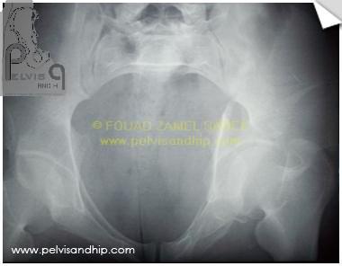
Although an acetabular fracture is not an indication for an inlet view, with inward displacement such as these it is sometimes useful to appreciate dispalcement, reduction and fixation on this view.
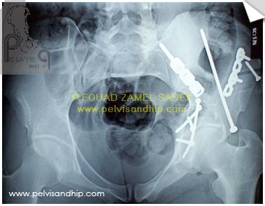
The reconstruction of the brim as well as the path of the LC II screw can be best visualized on this view.

If you feel like posting comments, enquiries or questions please click here.